Jump to section
Knowledge Map
Physiology
Overview
Management
Management
Prone Positioning
Neuromuscular Blockade
Evidence
Acute Respiratory Distress Syndrome
Acute, diffuse inflammatory lung injury causing hypoxemia and bilateral infiltrates not fully explained by cardiac failure or fluid overload.
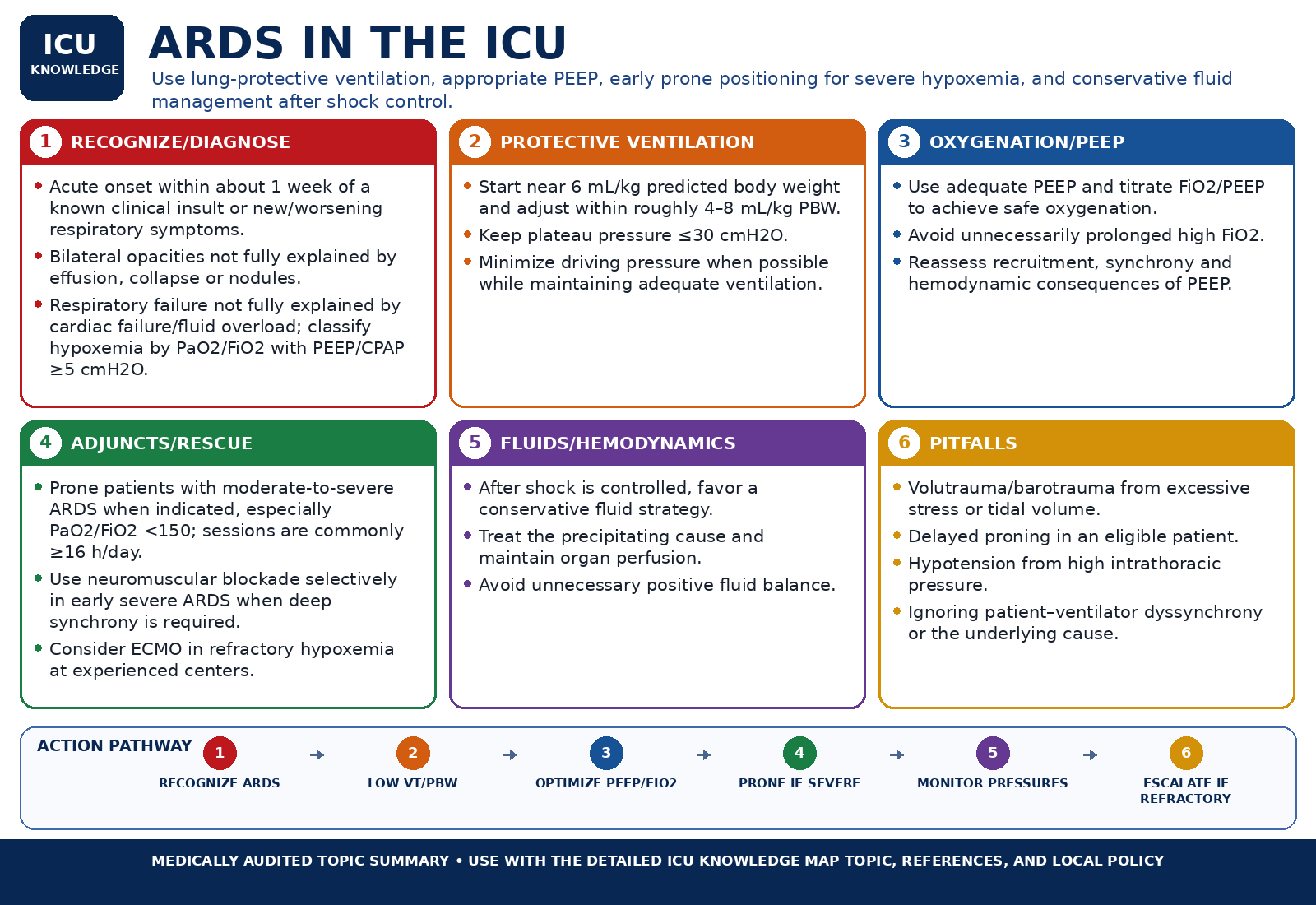
At-a-glance visual summary
Medical audit passed · 03 Sep 2026Use this as a rapid orientation only. The detailed topic text, references, and local protocols remain the clinical source of truth.
ARDS in the ICU — Audited At-a-Glance Summary
Clinical Orientation
Acute, diffuse inflammatory lung injury causing hypoxemia and bilateral infiltrates not fully explained by cardiac failure or fluid overload.
A common final pathway for many different insults to the alveolar-capillary membrane.
Acute dyspnea and hypoxemia in a patient with a recognized risk factor, evolving over hours to days.
Supportive care built around lung-protective ventilation, with escalation based on severity.
Both the disease and its treatment carry complication risk.
Overview
A common final pathway for many different insults to the alveolar-capillary membrane.
ARDS is not a single disease but a shared endpoint: pneumonia, sepsis, aspiration, trauma, transfusion, and pancreatitis can all injure the alveolar-capillary membrane in the same way, producing acute hypoxemic respiratory failure with a common radiographic and physiologic picture regardless of what triggered it.
It is one of the more common reasons for mechanical ventilation in the ICU, and its severity — and the risk that goes with it — varies considerably from patient to patient. There is no single drug or procedure that reverses the underlying injury; management is almost entirely about protecting the lung from additional, iatrogenic injury while the triggering illness is treated, which is why the Management section below is the largest part of this topic.
Definition
The Berlin Definition frames ARDS by timing, imaging, the origin of edema, and oxygenation.
ARDS: Histopathological Progression from Exudative Flooding to Pulmonary Fibrosis
ARDS pathology — overlapping injury, repair, fibroproliferation and fibrosis rather than rigid calendar stages.
Pathophysiological Sequence (Layer 2)
ARDS is defined by four elements that must all be present together, rather than by any single lab value: an acute timeline, characteristic imaging, exclusion of a purely cardiac cause, and a defined degree of hypoxemia measured under standardized ventilator conditions.
| Timing | Onset within 1 week of a known clinical insult or new/worsening respiratory symptoms. |
|---|---|
| Imaging | Bilateral opacities on chest X-ray or CT, not fully explained by effusions, lobar collapse, or nodules. |
| Origin of edema | Respiratory failure not fully explained by cardiac failure or fluid overload; requires objective assessment (e.g. echocardiography) if no clear risk factor is present. |
| Oxygenation | Graded by PaO2/FiO2 ratio measured at PEEP ≥ 5 cmH2O (see Severity). |
ARDS is a clinical syndrome with many possible triggers — pneumonia, sepsis, aspiration, trauma, transfusion, and pancreatitis are among the most common — rather than a single disease with one cause.
Guideline note
The table above is the 2012 Berlin Definition, which remains the framework nearly every major ARDS trial (ARMA through DEXA-ARDS) was designed and interpreted against. A 2024 Global Definition of ARDS built on Berlin rather than replacing its logic — see below for what it changes.The 2024 Global Definition of ARDS extends the same four-element framework to patients who are not yet intubated and to settings without arterial blood gas or standardized ventilator access. It adds a non-intubated ARDS category (PaO2/FiO2 ≤ 300, or SpO2/FiO2 ≤ 315 when SpO2 ≤ 97%, on high-flow nasal oxygen ≥ 30 L/min or noninvasive ventilation/CPAP ≥ 5 cmH2O), permits SpO2/FiO2 as an alternative to PaO2/FiO2 throughout, and adds a resource-variable category for settings without reliable PEEP delivery or blood gas access (SpO2/FiO2 ≤ 315, with neither a minimum PEEP nor a flow-rate requirement). Bilateral opacities may be assessed by lung ultrasound as well as chest X-ray or CT under the 2024 definition.
Caution
This content uses Berlin terminology and PaO2/FiO2 thresholds as the primary framework, since that is what the cited trials and the 2017/2023 ventilation guidelines were built around, and adds the 2024 Global Definition's SpO2/FiO2 and non-intubated criteria alongside it — the two are not in conflict, but a chart or trial that says "ARDS" without specifying which definition was applied should be read as Berlin unless stated otherwise.Physiology
Diffuse alveolar damage produces shunt physiology and a heterogeneous, poorly compliant lung.
ARDS Pathophysiology: Alveolar-Capillary Barrier Disruption & Shunt Heterogeneity
Textbook anatomical comparison: Intact alveolar-capillary barrier vs diffuse alveolar damage in ARDS.
Pathophysiological Sequence (Layer 2)
The pathologic correlate of early ARDS is diffuse alveolar damage: capillary leak floods alveoli with protein-rich edema fluid, surfactant function is disrupted, and alveoli collapse or fill with exudate. The result is a lung that is not uniformly stiff but heterogeneous — some regions remain relatively normal while others are fully consolidated or collapsed, so a single tidal volume is distributed disproportionately into the smaller aerated portion of the lung (sometimes described as a functionally 'baby lung').
Two physiologic consequences follow directly. First, perfused-but-unventilated alveoli create intrapulmonary shunt, producing hypoxemia that resists supplemental oxygen and instead requires alveolar recruitment. Second, because the aerated lung volume is small, a tidal volume that seems modest relative to total body size can still overdistend the aerated regions, causing ventilator-induced lung injury — the physiologic rationale for low tidal volume ventilation.
Guideline note
See the Physiology section for two standalone concepts this topic depends on: the Oxygen Delivery Equation and Ventilation-Perfusion Mismatch and Shunt.Clinical Presentation
Acute dyspnea and hypoxemia in a patient with a recognized risk factor, evolving over hours to days.
- Acute onset of dyspnea and hypoxemia, typically within a week of a known risk factor (pneumonia, sepsis, aspiration, trauma, transfusion, pancreatitis).
- Tachypnea and increased work of breathing; findings can be subtle early and progress quickly.
- Diffuse crackles on auscultation, though the exam can be relatively unremarkable in early or mild disease.
- No single exam finding is specific to ARDS — the presentation overlaps substantially with its differential diagnoses (see below).
Caution
The absence of volume-overload signs (jugular venous distension, peripheral edema, an S3) argues against a purely cardiogenic cause but does not exclude ARDS coexisting with a cardiac contribution — the two are not mutually exclusive.Diagnosis
Guideline
Diagnosis is clinical: apply the Berlin criteria and actively exclude a cardiac cause.
- Confirm the timing criterion: new or worsening respiratory symptoms within 1 week of a recognized risk factor.
- Review chest imaging for bilateral opacities not better explained by effusion, collapse, or nodules.
- Actively assess for a cardiac cause — clinical exam, BNP/NT-proBNP, and echocardiography when there is no obvious risk factor for ARDS or when the picture is ambiguous.
- Calculate the PaO2/FiO2 ratio at PEEP ≥ 5 cmH2O to grade severity once the syndromic diagnosis is met.
Caution
Volume overload and cardiogenic edema are the most common diagnostic pitfalls — they can produce bilateral opacities and hypoxemia that superficially mimic ARDS. Objective assessment of cardiac function is required whenever the cause is not obvious.Severity
Guideline
Berlin severity is graded by PaO2/FiO2 ratio at PEEP ≥ 5 cmH2O, and should be reassessed after optimization.
| Mild | PaO2/FiO2 200-300 (SpO2/FiO2 235-315 under the 2024 Global Definition) |
|---|---|
| Moderate | PaO2/FiO2 100-200 (SpO2/FiO2 148-235) |
| Severe | PaO2/FiO2 ≤ 100 (SpO2/FiO2 ≤ 148) |
Severity is not fixed at diagnosis — a patient classified as moderate on presentation may reclassify as severe or mild after PEEP and FiO2 are optimized over the first hours, and re-grading at that point is what should drive decisions like prone positioning.
Guideline note
The management evidence below (low tidal volume, PEEP, prone positioning, neuromuscular blockade) was generated using Berlin/PaO2-FiO2-defined severity. SpO2/FiO2 bands from the 2024 Global Definition are a validated proxy, not a separately trial-tested severity scale — treat them as approximate when they diverge from a directly measured PaO2/FiO2.PaO2/FiO2 Ratio
Grades hypoxemia severity using the 2012 Berlin ARDS oxygenation bands when PaO2/FiO2 is measured with PEEP/CPAP ≥5 cmH2O. The newer Global ARDS Definition also recognizes nonintubated ARDS on HFNO ≥30 L/min and additional pathways, so this calculator should not be used as the sole modern ARDS diagnostic tool.
Formula
P/F ratio = PaO2 / FiO2
Result
Enter all values to see the result.
Limitations
- The severity bands shown here are the Berlin 2012 PaO2/FiO2 categories and assume PEEP/CPAP ≥5 cmH2O. The Global ARDS Definition expands diagnosis to selected nonintubated patients (including HFNO ≥30 L/min), so absence of Berlin-compatible PEEP does not by itself exclude ARDS under the newer framework.
- Does not itself establish an ARDS diagnosis; the Berlin definition's timing, imaging, and cardiac-exclusion criteria must also be met.
- FiO2 is often only approximately known on non-invasive or high-flow oxygen devices, which limits precision outside invasive mechanical ventilation.
References
- ARDS Definition Task Force. Berlin Definition of ARDS. JAMA 2012.
- Matthay MA, et al. A New Global Definition of Acute Respiratory Distress Syndrome. Am J Respir Crit Care Med. 2024;209:37-47.
Differential Diagnosis
Conditions that can mimic the Berlin criteria and should be considered before settling on ARDS.
- Cardiogenic pulmonary edema — the most common mimic; look for volume-overload signs and consider echocardiography or a BNP.
- Diffuse alveolar hemorrhage — consider with hemoptysis, a falling hematocrit, or a known bleeding diathesis or vasculitis.
- Acute eosinophilic pneumonia — a rapidly progressive presentation, sometimes with peripheral or bronchoalveolar-lavage eosinophilia.
- Acute interstitial pneumonia (Hamman-Rich syndrome) — a diffuse alveolar damage pattern without an identifiable ARDS risk factor.
- Drug-induced lung injury — review the medication list for known culprits.
- Transfusion-related acute lung injury (TRALI) — onset within hours of a blood product transfusion.
Management
Guideline
Supportive care built around lung-protective ventilation, with escalation based on severity.
There is no therapy that reverses the underlying alveolar injury of ARDS; management is supportive, aimed at maintaining gas exchange while avoiding ventilator-induced lung injury until the lung recovers or the precipitating illness resolves. Treating the underlying cause (e.g. source control for sepsis) runs in parallel and is equally essential.
Lung-Protective Ventilation
GuidelineLow tidal volume with a plateau pressure ceiling is the foundational intervention in ARDS.
Driving Pressure – Compliance Relationship: ΔP = VT / Crs in Normal vs ARDS Lungs
AI-generated educational illustration: Driving pressure and compliance relationship — ΔP = VT / Crs comparison in normal compliance (50 mL/cmH2O) vs stiff ARDS lung (20 mL/cmH2O).
Pathophysiological Sequence (Layer 2)
- Target tidal volume approximately 6 mL/kg predicted body weight (range 4-8 mL/kg).
- Keep plateau pressure ≤ 30 cmH2O, reducing tidal volume further if needed to stay under this ceiling.
- Applies broadly across ARDS severity, not only to severe disease.
Trial note
Grounded in the ARMA (ARDSNet) trial, which showed a mortality reduction with 6 mL/kg vs 12 mL/kg ventilation.PEEP Titration
GuidelineHigher PEEP without a recruitment maneuver is favored in moderate-to-severe ARDS; driving pressure helps judge whether it's helping or overdistending the lung.
PEEP Titration: Alveolar Recruitment vs Overdistension & Hemodynamics
AI-generated educational illustration: PEEP titration spectrum — Alveolar collapse, optimal recruitment, overdistension, and preload reduction.
Pathophysiological Sequence (Layer 2)
PEEP recruits collapsed alveoli and reduces shunt, but excessive PEEP can overdistend already-aerated regions and worsen driving pressure. Current guidance conditionally favors a higher-PEEP strategy without a formal recruitment maneuver over a lower-PEEP strategy in moderate-to-severe ARDS, titrated against oxygenation and mechanics rather than fixed at one number for every patient.
Trial note
ALVEOLI found no difference in mortality or ventilator-free days between fixed higher- and lower-PEEP protocols in an unselected ALI/ARDS population — the guideline's preference for higher PEEP rests on later severity-stratified analyses, not on ALVEOLI's own null result.Caution
The 2023 ESICM guideline strongly recommends against prolonged, aggressive lung recruitment maneuvers in moderate-to-severe ARDS — see Landmark Trials for the ART trial, which found higher mortality with an aggressive recruitment-plus-titrated-PEEP protocol.Driving Pressure
Plateau pressure minus PEEP — a marker of lung-protective ventilation beyond tidal volume alone.
Formula
Driving pressure = Plateau pressure − PEEP
Result
Enter all values to see the result.
Limitations
- Requires a valid plateau pressure measured during an end-inspiratory pause with no active patient effort — spontaneous breathing effort invalidates the measurement.
- The mortality association comes from retrospective analyses of trial data, not a randomized trial of a driving-pressure-targeted strategy itself.
- Doesn't separate lung from chest wall contribution — see transpulmonary pressure for that distinction.
References
- Amato MB, et al. Driving Pressure and Survival in the Acute Respiratory Distress Syndrome. NEJM 2015.
Fluid Management
RCTAfter initial resuscitation, avoid unnecessary positive balance and transition toward conservative or active fluid removal when perfusion and congestion assessment support it.
- During active shock resuscitation, restoring perfusion takes priority; do not force a negative balance while the patient remains clearly preload responsive or under-resuscitated.
- After the acute resuscitation phase, stop fluids that no longer have an indication and use a conservative strategy; active diuresis or ultrafiltration should be guided by congestion, renal function, hemodynamics and tolerance rather than by a fixed net-balance target.
- Ongoing low-dose vasopressor use is not by itself a universal prohibition on deresuscitation, but it increases the need for repeated perfusion and fluid-responsiveness assessment.
- FACTT supports more ventilator-free days with a conservative strategy after shock stabilization; it did not demonstrate a mortality benefit.
Trial note
FACTT demonstrated more ventilator-free days with conservative fluid management after stabilization, while contemporary sepsis guidance emphasizes individualized de-resuscitation rather than a mandatory timing or balance formula.Prone Positioning
RCTUse early prolonged prone positioning in appropriate moderate-to-severe ARDS, particularly when PaO2/FiO2 remains <150 despite an optimized lung-protective strategy.
- For intubated ARDS with PaO2/FiO2 <150 despite lung-protective ventilation and appropriate PEEP, prone positioning should be instituted early when no overriding safety issue exists.
- Use prolonged sessions, generally at least 16 consecutive hours, with a trained team and explicit airway/line rescue planning.
- Do not stop a safe prone session solely because oxygenation fails to improve immediately; the benefit is not limited to immediate oxygenation responders.
Indications
Severe, persistent hypoxemia despite an optimized lung-protective strategy.
PaO2/FiO2 below 150 that persists despite lung-protective ventilation, adequate PEEP, and a reasonable trial of optimization — this is the population in which the mortality benefit has actually been demonstrated, not a general therapy for any hypoxemic patient.
Patient Selection
Confirm the airway, lines, and hemodynamics can tolerate safe turning before proceeding.
Before turning, confirm the airway is secure, central and arterial lines are unlikely to dislodge, and the patient is hemodynamically stable enough to tolerate the maneuver. Obesity alone is not a contraindication but does require extra staff and appropriate equipment.
Duration
Sessions of at least 16 hours, reassessed daily against oxygenation once supine.
Each proning session should last at least 16 consecutive hours. Oxygenation is reassessed after a period back supine; proning is generally stopped once oxygenation is sustained (PaO2/FiO2 above roughly 150) supine with modest PEEP and FiO2 across multiple attempts.
Contraindications
Separate true barriers to turning from relative hazards that can often be managed with specialist planning.
- Unstable spinal injury is a major contraindication unless a specialist team determines turning can be performed safely.
- Open abdomen, recent abdominal/thoracic surgery, elevated intracranial pressure, facial/airway surgery and major hemodynamic instability are not uniform absolute contraindications; weigh the specific anatomy, devices and physiologic risk with the relevant specialist team.
- Obesity and pregnancy are not automatic exclusions but require adequate staffing, positioning equipment and pressure-point/abdominal protection.
Complications
Mostly positioning-related, not a consequence of the physiology itself.
- Facial and airway edema.
- Pressure injuries at the face, chest, and knees.
- Transient desaturation or hemodynamic changes during the turn itself.
- Endotracheal tube or line displacement.
Neuromuscular Blockade
GuidelineSelective rather than automatic: SCCM 2026 conditionally supports NMBA when PaO2/FiO2 <150 and hypoxemia or ventilation targets remain unacceptable despite sedation.
Routine paralysis of every patient with moderate-to-severe ARDS is not supported. SCCM 2026 conditionally suggests an NMBA for adults with ARDS and PaO2/FiO2 <150 who remain persistently hypoxemic and/or are not achieving mechanical-ventilation targets despite adequate analgesia and sedation. Patient-ventilator dyssynchrony, injurious respiratory effort and inability to deliver lung-protective ventilation are common practical reasons to use a short course.
Guideline note
SCCM 2026: use NMBA selectively in PaO2/FiO2 <150 with persistent hypoxemia and/or failure to achieve ventilation targets on sedation (conditional, low-certainty). Prone positioning alone does not mandate NMBA use.Controversy
ACURASYS suggested benefit under deep-sedation-era care, whereas ROSE found no mortality benefit compared with a lighter-sedation strategy. The modern recommendation is conditional and phenotype/ventilation-target driven, not a universal ARDS bundle item.Corticosteroids
RCTATS 2024 conditionally suggests corticosteroids for ARDS, but the optimal agent, dose, timing and duration remain uncertain.
- ATS 2024 conditionally suggests corticosteroids for adults with ARDS (moderate certainty of evidence).
- Do not convert that recommendation into one mandatory dexamethasone or methylprednisolone regimen: trials used heterogeneous drugs, doses, timing and durations, and the guideline did not establish a single preferred protocol.
- Consider infection, immunosuppression, neuromuscular weakness, hyperglycemia and the timing/etiology of ARDS when individualizing therapy.
Trial note
DEXA-ARDS is an important positive trial in established moderate-to-severe non-COVID ARDS, but the ATS 2024 recommendation is based on the broader corticosteroid evidence base rather than on that trial alone.Rescue Therapies
RCTATS 2024 conditionally supports VV-ECMO for selected severe ARDS after evidence-based conventional management has been optimized.
For selected patients with severe ARDS and refractory hypoxemia or injurious ventilatory requirements despite optimized lung-protective ventilation, PEEP and prone positioning, ATS 2024 conditionally suggests venovenous ECMO (low-certainty evidence). Selection should account for reversibility, duration of injurious ventilation, comorbidity, contraindications, center expertise and an explicit exit strategy; ECMO is not an automatic next step based on a single PaO2/FiO2 value.
Monitoring
What to trend day to day: ventilator mechanics, gas exchange, hemodynamics, and readiness to wean.
- Ventilator mechanics: plateau pressure, driving pressure, and compliance, trended over time rather than checked once.
- Gas exchange: arterial blood gases and PaO2/FiO2, reassessed after any change to PEEP or FiO2.
- Hemodynamics: particularly important around proning and high-PEEP titration, both of which can affect venous return and right ventricular afterload.
- Sedation and analgesia depth, especially if neuromuscular blockade is in use.
- Daily assessment for readiness to reduce support once oxygenation and mechanics are improving.
Caution
Specific monitoring targets and protocols vary by institution — confirm against local ICU protocol rather than treating the items above as fixed thresholds.Complications
Both the disease and its treatment carry complication risk.
- Barotrauma, including pneumothorax, from high airway pressures or underlying lung fragility.
- Ventilator-associated pneumonia from prolonged intubation.
- ICU-acquired weakness, compounded by immobility, deep sedation, and neuromuscular blockade.
- Fibroproliferative changes in a subset of patients with prolonged ARDS, contributing to persistent restrictive physiology.
- Right ventricular strain from hypoxic vasoconstriction and elevated intrathoracic pressure at high PEEP.
Guidelines
Guideline
Current guidance is anchored by ATS 2024 and ESICM 2023, with SCCM 2026 refining NMBA selection; earlier ATS/ESICM/SCCM recommendations remain relevant where retained.
ATS 2024 conditionally suggests corticosteroids, VV-ECMO in selected severe ARDS, neuromuscular blockade in selected early severe ARDS, and higher PEEP without prolonged recruitment maneuvers in moderate-to-severe ARDS; it strongly recommends against prolonged recruitment maneuvers. SCCM 2026 further narrows NMBA selection to PaO2/FiO2 <150 with persistent hypoxemia and/or inability to achieve ventilation targets despite sedation. Low tidal-volume ventilation with plateau pressure limitation and prolonged prone positioning remain core evidence-based therapy.
Landmark Trials
RCT
Nine trials that define current ARDS management, spanning ventilation, PEEP strategy, fluid management, positioning, sedation, steroids, and rescue therapy.
- ARMA (ARDSNet, 2000) — established low tidal volume ventilation.
- ALVEOLI (2004) — found no difference between fixed higher- and lower-PEEP protocols in an unselected population.
- FACTT (2006) — established conservative fluid management after shock resolution shortens ventilator time, without a mortality difference.
- ACURASYS (2010) — reported an adjusted mortality benefit with early neuromuscular blockade.
- PROSEVA (2013) — established early, prolonged prone positioning in severe ARDS.
- ART (2017) — found higher mortality with an aggressive lung recruitment and titrated-PEEP strategy compared with a conventional low-PEEP approach.
- EOLIA (2018) — tested early ECMO in very severe ARDS; primary endpoint not significant, interpretation complicated by crossover.
- ROSE (2019) — found no mortality benefit with routine early neuromuscular blockade, contradicting ACURASYS.
- DEXA-ARDS (2020) — a single-country trial reporting a large mortality benefit with early dexamethasone in moderate-to-severe ARDS, not yet confirmatory-trial-replicated.
Recent Evidence
Meta-analysis of neuromuscular blockade trials illustrates why pooling trials with different comparator strategies is difficult.
A pooled analysis of neuromuscular blockade trials in ARDS found no clear overall mortality benefit, with the estimate highly sensitive to the sedation strategy used in each trial's comparator arm — a reminder that meta-analysis cannot fully resolve a question when the underlying trials are not testing quite the same clinical strategy.
Controversies
RCT
The main controversies now concern whom to paralyze, which corticosteroid regimen to use, how to individualize PEEP and when to refer for ECMO.
Controversy
Neuromuscular blockade should no longer be framed as simply 'routine' versus 'never.' SCCM 2026 conditionally favors it in PaO2/FiO2 <150 when hypoxemia persists or lung-protective ventilation targets cannot be achieved despite sedation, while acknowledging low-certainty evidence.ATS 2024 also moved corticosteroids and selected VV-ECMO into conditional guideline-supported options, while leaving important implementation uncertainty: steroid regimen and timing, individualized PEEP/recruitability, and ECMO selection should be tailored rather than reduced to fixed formulas.
Clinical Problems Where This Topic is Actionable (4)
Start from the patient presentation and determine immediate bedside priorities, differentials, and triggers.
Sudden Hypoxemia
Bedside triage of acute desaturation: signal verification, immediate danger screening (DOPE / tension pneumothorax), oxygen escalation ladder, and mechanism-based differential.
Sudden Ventilator Deterioration & High Airway Pressures
Algorithmic troubleshooting of acute patient-ventilator collapse: the DOPE framework, peak vs plateau pressure differentiation, auto-PEEP evaluation, and manual bagging separation.
Acute Severe Respiratory Distress
Bedside management of the gasping, tachypneic patient: work-of-breathing assessment, accessory muscle use, P-SILI prevention, and timely intubation vs noninvasive support.
Immunocompromised Patient with Fever + Organ Failure
Bedside diagnostic framing and emergency management of acute fever, sepsis, respiratory failure, or oncologic emergency in an immunocompromised host.
Clinical Decision Pathways (7)
Step-by-step ICU protocols: Trigger → Assess → Decision → Intervene → Reassess → Escalate.
Mechanical Ventilation Initiation Workflow
From post-intubation verification and lung-protective settings to mechanics optimization.
Mechanical Ventilation Liberation & Weaning
From readiness screening and spontaneous breathing trials to extubation and post-extubation support.
Prone Positioning Protocol for Severe ARDS
From eligibility screening and team preparation to 16-hour proning cycles and response evaluation.
Noninvasive Respiratory Support Escalation & Intubation Decision
From initial noninvasive modality selection to ROX index tracking and timely intubation triggers.
New ARDS Diagnosis — First 6 Hours
From recognition to lung-protective ventilation and severity-driven escalation.
Acute Hypoxemia
From confirming true hypoxemia to identifying the underlying mechanism.
Neutropenic & Immunocompromised Sepsis
Immediate 1-hour empiric antipseudomonal therapy, host-specific diagnostic pathway, and oncologic emergency escalation.
Clinical Calculators (5)
Formula-based ICU calculators to guide severity stratification and therapeutic titration.
PaO2/FiO2 Ratio
Grades hypoxemia severity using the 2012 Berlin ARDS oxygenation bands when PaO2/FiO2 is measured with PEEP/CPAP ≥5 cmH2O. The newer Global ARDS Definition also recognizes nonintubated ARDS on HFNO ≥30 L/min and additional pathways, so this calculator should not be used as the sole modern ARDS diagnostic tool.
Driving Pressure
Plateau pressure minus PEEP — a marker of lung-protective ventilation beyond tidal volume alone.
Static Compliance
Tidal volume delivered per unit of driving pressure — a bedside marker of how stiff the lung and chest wall are.
Ideal Body Weight
Predicted body weight from height and sex (Devine formula) — the basis for lung-protective tidal volume dosing.
Tidal Volume by Predicted Body Weight
Target tidal volume for lung-protective ventilation, dosed to predicted body weight rather than actual weight.
Evidence Timeline
Assess Your Knowledge
Master this topic with high-yield spaced-repetition flashcards and board-style clinical reasoning questions.
Topic Flashcards
Review definitions, pathophysiology, diagnostic criteria, key thresholds, and trial findings.
Practice Questions
Test your clinical decision-making with board-style clinical vignettes and detailed rationale.
Evidence status
Review metadata not yet recorded.
Status, review dates and guideline versions appear here once editorial metadata is authored for this content.
Sources & Provenance
Foundation
✓ Verified textbook sourceIrwin & Rippe's Intensive Care Medicine, 9th Edition, 2023
Guidelines
✓ 8 verifiedATS/ESICM/SCCM — Mechanical Ventilation in Adult Patients with ARDS
American Thoracic Society / European Society of Intensive Care Medicine / Society of Critical Care Medicine, 2017
ATS/ESICM/SCCM — Mechanical Ventilation in Adult Patients with ARDS
American Thoracic Society / European Society of Intensive Care Medicine / Society of Critical Care Medicine, 2017
ATS/ESICM/SCCM — Mechanical Ventilation in Adult Patients with ARDS
American Thoracic Society / European Society of Intensive Care Medicine / Society of Critical Care Medicine, 2017
ESICM — Guidelines on Acute Respiratory Distress Syndrome: Definition, Phenotyping and Respiratory Support Strategies
European Society of Intensive Care Medicine, 2023
ESICM — Guidelines on Acute Respiratory Distress Syndrome: Definition, Phenotyping and Respiratory Support Strategies
European Society of Intensive Care Medicine, 2023
SCCM — Guidelines for the Administration of Neuromuscular Blockade in Adults With ARDS
Society of Critical Care Medicine, 2026
ESICM — Guidelines on Acute Respiratory Distress Syndrome: Definition, Phenotyping and Respiratory Support Strategies
European Society of Intensive Care Medicine, 2023
ATS — An Update on Management of Adult Patients with Acute Respiratory Distress Syndrome
American Thoracic Society, 2024
Primary Evidence
✓ 9 verifiedARMA (ARDSNet)
New England Journal of Medicine, 2000
PROSEVA
New England Journal of Medicine, 2013
ACURASYS
New England Journal of Medicine, 2010
ROSE
New England Journal of Medicine, 2019
EOLIA
New England Journal of Medicine, 2018
ART
JAMA, 2017
ALVEOLI
New England Journal of Medicine, 2004
FACTT
New England Journal of Medicine, 2006
DEXA-ARDS
The Lancet Respiratory Medicine, 2020
Synthesis
✓ 1 verifiedCurrent Status
Current through the ATS 2024 ARDS management update, ESICM 2023 ARDS guideline, SCCM 2026 neuromuscular-blockade guideline, and the 2024 Global Definition of ARDS. Low tidal-volume ventilation and prolonged prone positioning remain core therapy; corticosteroids, selected VV-ECMO, selected NMBA use and higher PEEP without prolonged recruitment maneuvers are guideline-supported with conditional patient selection.
Other References
- ARDS Definition Task Force. Berlin Definition of ARDS. JAMA 2012.
- Matthay MA, et al. A New Global Definition of Acute Respiratory Distress Syndrome. Am J Respir Crit Care Med. 2024;209(1):37-47.
- Qadir N, et al. An Update on Management of Adult Patients with Acute Respiratory Distress Syndrome: An Official ATS Clinical Practice Guideline. Am J Respir Crit Care Med. 2024;209:24-36. doi:10.1164/rccm.202311-2011ST.
- Erstad BL, Sarwal A, et al. SCCM Guidelines for the Administration of Neuromuscular Blockade in Adults With ARDS. Crit Care Med. 2026;54:634-643.
Continue Learning
Cisatracurium
Non-Depolarizing Benzylisoquinolinium NMBA
Dexamethasone
Long-Acting Synthetic Fluorinated Glucocorticoid (High Anti-Inflammatory Potency) with Negligible Mineralocorticoid Activity
Severe ARDS Ventilator Strategy
Lung-protective ventilation, prone positioning, PEEP, and selective neuromuscular blockade
ARDS: Driving Pressure
Using respiratory-system mechanics without turning one marker into a rigid target
Sudden Hypoxemia
Bedside triage of acute desaturation: signal verification, immediate danger screening (DOPE / tension pneumothorax), oxygen escalation ladder, and mechanism-based differential.
Sudden Ventilator Deterioration & High Airway Pressures
Algorithmic troubleshooting of acute patient-ventilator collapse: the DOPE framework, peak vs plateau pressure differentiation, auto-PEEP evaluation, and manual bagging separation.
What Next? — Continue Your Clinical Workflow
Mechanical Ventilation Initiation Workflow
From post-intubation verification and lung-protective settings to mechanics optimization.
Sudden Hypoxemia
Bedside triage of acute desaturation: signal verification, immediate danger screening (DOPE / tension pneumothorax), oxygen escalation ladder, and mechanism-based differential.
Test Clinical Reasoning
Answer scenario-based clinical questions with detailed rationales and trial links.
Respiratory Failure: Basic Principles
Core taxonomy (Types I–IV), alveolar gas exchange physiology, work-of-breathing mechanics, and escalation thresholds in acute respiratory failure.
PaO2/FiO2 Ratio
Grades hypoxemia severity using the 2012 Berlin ARDS oxygenation bands when PaO2/FiO2 is measured with PEEP/CPAP ≥5 cmH2O. The newer Global ARDS Definition also recognizes nonintubated ARDS on HFNO ≥30 L/min and additional pathways, so this calculator should not be used as the sole modern ARDS diagnostic tool.
Explore Knowledge Map
Explore the entire critical care network and inspect cross-system relationships.