ICU ECG Atlas
Look at the ECG, choose the diagnosis, then choose why. No free-text interpretation is required.
Standard 3×4 · 2.5 s/lead · 10 s rhythm strip · 25 mm/s · 10 mm/mV
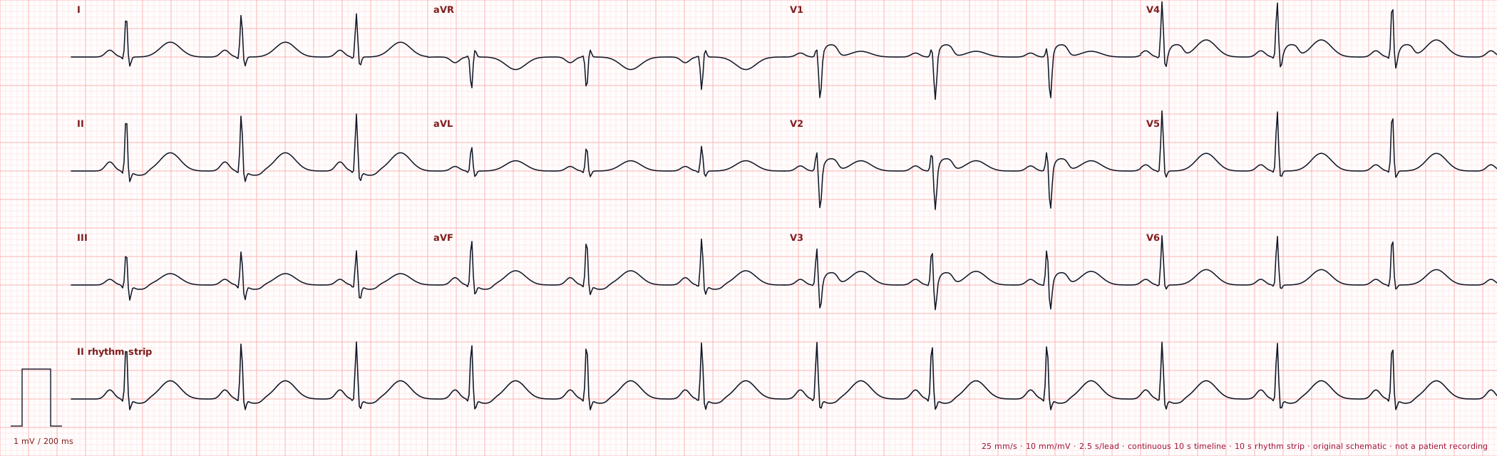
Calibrated original schematic ECGs · standard 3×4 lead layout
8
Modules
47
Choice cases
2
Choices per case
0
Free-text fields
0/47
Cases attempted
0
Attempts
0%
Full-score accuracy
Access model
Foundations + selected cases are available in Guest Preview
Advanced modules, Compare mode, and the balanced 20-case exam require a free signed-in account.
Curriculum
The trimmed ICU-focused curriculum you approved.
Module 1
ECG Foundations
Recognize normal sinus patterns, rate changes, and common artifact before ICU emergencies.
Module 2
ICU Tachyarrhythmias
High-yield narrow- and wide-complex tachyarrhythmias, including unstable ventricular rhythms.
Module 3
Bradyarrhythmias
Sinus and junctional bradycardia plus the AV block patterns that matter in the ICU.
Module 4
Ischemia & Infarction
Recognize major infarction territories and high-risk diffuse ischemic patterns.
Module 5
Electrolyte & Metabolic ECGs
Pattern recognition for common ICU electrolyte emergencies and hypothermia.
Module 6
Toxicology ECG
Recognize ECG patterns that may accompany cardiotoxic exposures without over-calling the toxin from ECG alone.
Module 7
Pulmonary / Right Heart
Selected ECG patterns of acute right-heart strain that may occur with pulmonary embolism.
Module 8
Paced Rhythm
Recognize paced rhythm morphology without turning this into a device troubleshooting course.
Practice
Diagnosis + why
Case names stay neutral until submission and option order is shuffled deterministically.
Compare
Look-alike patterns
AF vs flutter, Mobitz I vs II, VT vs pacing, ischemic patterns, and more.
Exam
Balanced 20-ECG challenge
All eight modules are represented and the answer key is graded server-side.
ECG sourcing & safety
The current traces are original calibrated schematic teaching ECGs built inside ICU Knowledge Map rather than copied from a textbook or teaching website. They use the conventional 3×4 lead arrangement, 25 mm/s timing, and 10 mm/mV calibration for pattern-recognition education. They are not patient recordings and must not be used for patient diagnosis. The data model remains ready for attributed licensed real-signal additions such as PhysioNet.