Jump to section
Knowledge Map
Acute Kidney Injury in Critical Care
Sepsis-AKI microcirculatory decoupling, KDIGO staging, Furosemide Stress Test (FST), urine microscopy, and the AEIOU dialysis threshold framework.
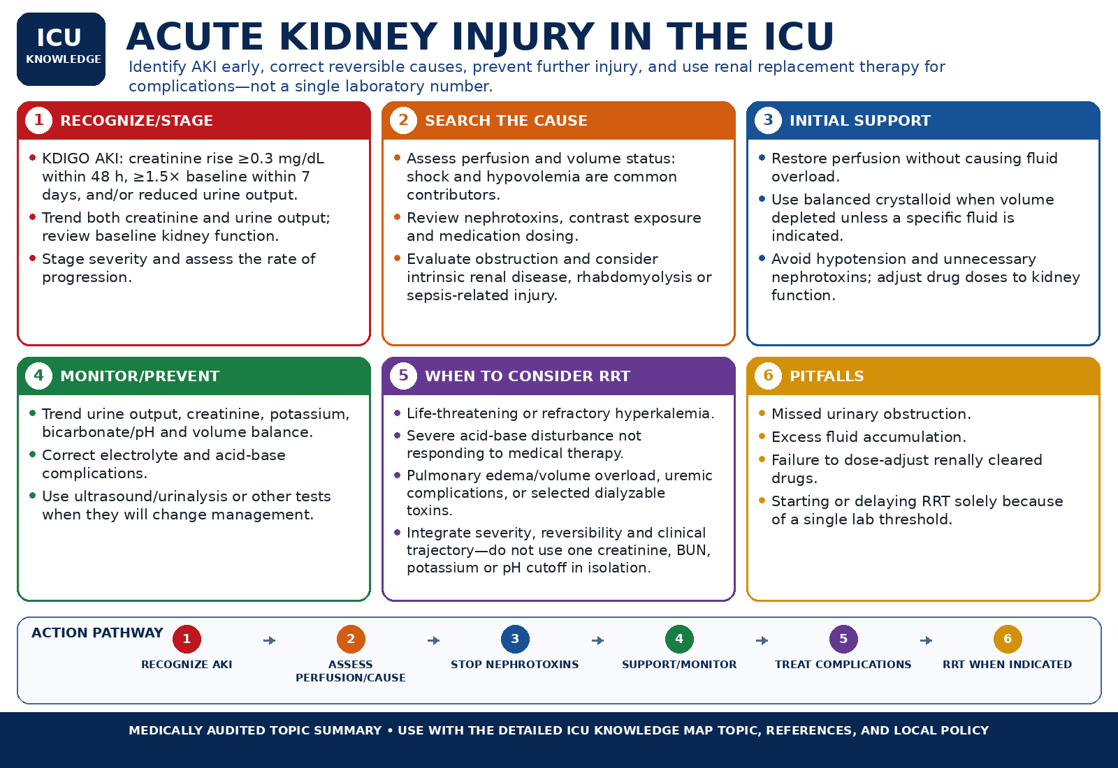
At-a-glance visual summary
Medical audit passed · 03 Sep 2026Use this as a rapid orientation only. The detailed topic text, references, and local protocols remain the clinical source of truth.
Acute Kidney Injury in the ICU — Audited At-a-Glance Summary
Clinical Orientation
Sepsis-AKI microcirculatory decoupling, KDIGO staging, Furosemide Stress Test (FST), urine microscopy, and the AEIOU dialysis threshold framework.
Acute Kidney Injury (AKI) is an abrupt decline in glomerular filtration and tubular function, staged by serum creatinine and urine output, carrying an ICU mortality of 30–50%.
Fractional excretion formulas, distinguishing prerenal azotemia from intrinsic ATN, and sediment microscopy findings.
Absolute emergency indications versus watchful waiting in non-emergent AKI.
Hyperkalemia, uremic bleeding, fluid overload-induced organ crosstalk, and transition to chronic kidney disease.
Overview
Acute Kidney Injury (AKI) is an abrupt decline in glomerular filtration and tubular function, staged by serum creatinine and urine output, carrying an ICU mortality of 30–50%.
Acute Kidney Injury: Ischemic & Nephrotoxic Acute Tubular Injury
Textbook anatomical comparison: Normal nephron function vs acute tubular injury with brush border loss, cast obstruction, and back-leak.
Pathophysiological Sequence (Layer 2)
Acute Kidney Injury (AKI) complicates up to 50% of all critically ill ICU patients and is an independent predictor of prolonged mechanical ventilation, multiorgan dysfunction, and hospital mortality. Sepsis is the single most common etiology (~50% of ICU AKI), followed by major surgery, cardiogenic shock, and nephrotoxin exposure.
Diagnostic evaluation requires rapid KDIGO staging, medication/hemodynamic review, urinalysis and urine microscopy, assessment for obstruction, and selective use of functional indices or the Furosemide Stress Test when they answer a specific question. FeNa/FeUrea and FST refine probability or prognosis; none independently distinguishes every prerenal versus intrinsic-AKI phenotype or mandates dialysis.
Differential Diagnosis of Acute Oliguria & Azotemia in Critical Care
Differentiate pre-renal hemodynamic azotemia, acute tubular necrosis (ATN), acute interstitial nephritis (AIN), and post-renal obstruction.
- Hemodynamic / pre-renal physiology vs tubular injury: traditional low FeNa (<1%) or low FeUrea (<35%) patterns can support sodium/urea avidity, while muddy-brown granular casts and renal tubular epithelial cells support tubular injury. These indices are not diagnostic cutoffs: sepsis, CKD, diuretics, glomerular disease, pigment injury and mixed shock frequently blur the classic pattern; integrate urine sediment, hemodynamics, obstruction assessment and the clinical trajectory.
- Abdominal Compartment Syndrome (ACS): High intra-abdominal pressure (IAP > 20 mmHg) compressing renal veins and parenchyma, causing sudden oliguria with elevated CVP; confirmed by bladder pressure measurement.
- Acute Interstitial Nephritis (AIN): Allergic drug reaction (antibiotics, PPIs, NSAIDs) presenting with sterile pyuria, WBC casts, eosinophiluria, and rash.
- Cardiorenal Syndrome Type 1: High central venous congestion (CVP > 15-20 mmHg) causing backward venous hypertension and drop in net renal perfusion pressure.
Definition & KDIGO Staging Matrix
Guideline
The 2012 KDIGO consensus staging system across Stage 1, Stage 2, and Stage 3 based on serum creatinine rise and duration of oliguria.
AKI is diagnosed when any ONE of the following three criteria is met:
| Diagnostic Criteria | 1. Serum creatinine increase by ≥ 0.3 mg/dL (≥ 26.5 umol/L) within 48 hours; OR 2. Serum creatinine increase to ≥ 1.5 times baseline within the prior 7 days; OR 3. Urine volume < 0.5 mL/kg/hour for ≥ 6 consecutive hours. |
|---|---|
| Stage 1 | Serum creatinine 1.5–1.9 times baseline OR increase ≥ 0.3 mg/dL; OR urine output < 0.5 mL/kg/hr for 6–12 hours. |
| Stage 2 | Serum creatinine 2.0–2.9 times baseline; OR urine output < 0.5 mL/kg/hr for ≥ 12 hours. |
| Stage 3 | Serum creatinine ≥ 3.0 times baseline OR increase to ≥ 4.0 mg/dL (≥ 353.6 umol/L) OR initiation of Renal Replacement Therapy; OR urine output < 0.3 mL/kg/hr for ≥ 24 hours OR anuria for ≥ 12 hours. |
Pathophysiology: Sepsis-AKI, Autoregulation, & Nephrotoxins
The sepsis-AKI microcirculatory paradigm, renal perfusion pressure (MAP - CVP), tubular mitochondrial hibernation, and nephrotoxicity mechanisms.
Glomerular Filtration Rate (GFR) & Renal Microvascular Autoregulation
AI-generated educational illustration: Glomerular filtration and renal autoregulation — afferent/efferent arteriolar tone and tubuloglomerular feedback.
Pathophysiological Sequence (Layer 2)
Glomerular Filtration Dynamics: Starling Forces & Arteriolar Hemodynamics
AI-generated educational illustration: Glomerular filtration dynamics — Starling pressures (Pgc, Pbs, πgc), net filtration pressure, and arteriolar resistance.
Pathophysiological Sequence (Layer 2)
Nephron Tubular Function: Solute & Water Handling Across Segments
AI-generated educational illustration: Nephron tubular function — Segmental map of solute reabsorption and secretion across PCT, Loop of Henle, DCT, and Collecting Duct.
Pathophysiological Sequence (Layer 2)
Loop Diuretic Physiology: Thick Ascending Limb NKCC2 Blockade & Electrolytes
AI-generated educational illustration: Loop diuretic physiology — Thick ascending limb NKCC2 cotransporter blockade, medullary gradient loss, and electrolyte excretion.
Pathophysiological Sequence (Layer 2)
Renal hemodynamic and cellular injury in the ICU is governed by complex microvascular and cellular mechanisms:
| 1. Renal Perfusion Pressure (RPP) | RPP = Mean Arterial Pressure (MAP) - Central Venous Pressure (CVP). In circulatory shock or venous congestion (VExUS grade 3), falling MAP and rising CVP drastically narrow net transglomerular filtration pressure, triggering oliguria. |
|---|---|
| 2. The Modern Sepsis-AKI Paradigm | Sepsis-AKI is NOT purely ischemic tubular necrosis. Human and experimental data show that sepsis causes profound renal microvascular flow heterogeneity, efferent arteriolar vasodilation (causing GFR collapse despite preserved global renal blood flow), pathogen-associated molecular pattern (PAMP) activation of TLR-4 receptors on tubular cells, and tubular cell mitochondrial shutdown (metabolic hibernation) to protect against apoptosis. |
| 3. Common ICU Nephrotoxin Mechanisms | • Vancomycin: Direct proximal tubular oxidative stress and cast formation. • Aminoglycosides: Megalin-receptor-mediated endocytosis into proximal tubule lysosomes causing phospholipidosis. • IV Iodinated Contrast: Medullary hypoxia from intense vasoconstriction + direct tubular cytotoxicity. • NSAIDs: Block vasodilatory prostaglandins -> afferent arteriolar vasoconstriction. • ACEi / ARBs: Block angiotensin II -> efferent arteriolar vasodilation. • Rhabdomyolysis: Free myoglobin precipitation with Tamm-Horsfall protein + heme-mediated lipid peroxidation. |
Diagnostic Protocol: FeNa, FeUrea, & Urine Microscopy
Guideline
Fractional excretion formulas, distinguishing prerenal azotemia from intrinsic ATN, and sediment microscopy findings.
| Fractional Excretion of Sodium (FeNa) | FeNa (%) = [(Urine Na × Serum Cr) / (Serum Na × Urine Cr)] × 100. Low or high values can support a physiology in the right context, but the classic <1% and >2% cutoffs are not diagnostic: sepsis, CKD, glomerular disease, pigment injury, contrast exposure, diuretics and mixed shock can all break the rule. |
|---|---|
| Fractional Excretion of Urea (FeUrea) | FeUrea (%) = [(Urine Urea × Serum Cr) / (Serum BUN × Urine Cr)] × 100. FeUrea may be less affected by loop diuretics than FeNa in some settings, but its diagnostic performance is also imperfect; <35% or >50% should not be used as stand-alone proof of prerenal azotemia or ATN. |
| Urine Sediment Microscopy ('Liquid Kidney Biopsy') | • Muddy brown granular casts and renal tubular epithelial (RTE) cells: strongly support acute tubular injury/ATN in the appropriate clinical context, but urine microscopy is not perfectly diagnostic in isolation. • White Blood Cell (WBC) Casts + Eosinophiluria: Suggests Acute Interstitial Nephritis (AIN). • Dysmorphic Red Blood Cells + RBC Casts: Indicates Acute Glomerulonephritis / Vasculitis. • Broad Waxy Casts: Reflects Chronic Kidney Disease / tubular dilation. |
Fractional Excretion of Sodium (FeNa)
Calculates FeNa as a context-dependent physiologic clue in AKI; it can support but does not by itself distinguish hemodynamic/prerenal physiology from intrinsic kidney injury.
Formula
FeNa = (Urine Na × Plasma Cr) / (Plasma Na × Urine Cr) × 100
Result
Enter all values to see the result.
Limitations
- Loop/thiazide diuretics can increase urine sodium and reduce FeNa's diagnostic usefulness; this makes interpretation less reliable rather than mathematically invalidating the calculation.
- Less reliable in patients with underlying chronic kidney disease, contrast-induced nephropathy, or certain glomerulonephritides, which can show a low FeNa despite intrinsic injury.
- A single value is a probabilistic aid, not a definitive diagnostic test — it should be interpreted alongside the full clinical picture, not in isolation.
References
- Standard fractional-excretion formula; use only as an adjunct to hemodynamics, urine sediment, obstruction assessment and clinical trajectory.
Fractional Excretion of Urea (FeUrea)
Calculates FeUrea, sometimes used as an adjunct when diuretics limit FeNa interpretation; FeUrea is also context-dependent and does not reliably distinguish prerenal physiology from intrinsic AKI by itself.
Formula
FeUrea = (Urine urea nitrogen × Plasma Cr) / (Plasma urea nitrogen × Urine Cr) × 100
Result
Enter all values to see the result.
Limitations
- FeUrea is not a validated 'fix' for FeNa after diuretics; its diagnostic performance is also imperfect in sepsis, CKD and mixed ICU physiology.
- Urea handling can also be affected by protein intake, catabolic state, and liver function, which can shift the result independent of the prerenal/intrinsic distinction.
- A single value is a probabilistic aid, not a definitive diagnostic test — it should be interpreted alongside the full clinical picture, not in isolation.
References
- Carvounis CP, et al. Significance of the fractional excretion of urea in the differential diagnosis of acute renal failure. Kidney Int. 2002.
Risk Stratification: The Furosemide Stress Test (FST)
RCT
Chawla et al. FST protocol for predicting progression to severe Stage 3 AKI and need for renal replacement therapy.
The Furosemide Stress Test (FST) is a validated functional challenge assessing the integrity of the loop of Henle and collecting duct in early AKI (Chawla et al., Crit Care 2013):
| FST Protocol | Patient must be euvolemic / adequately fluid resuscitated. Administer IV Furosemide: 1.0 mg/kg in loop-diuretic-naïve patients OR 1.5 mg/kg in patients with prior loop diuretic exposure. Measure hourly urine output. |
|---|---|
| FST Interpretation | Urine Output <200 mL in the 2 hours after furosemide is commonly used as a positive FST threshold. In the original derivation study, this threshold had about 87% sensitivity and 84% specificity for progression to KDIGO Stage 3 AKI. The FST is a risk-stratification tool: a response >200 mL/2 h suggests greater tubular functional reserve and a lower risk of severe AKI progression, but it does not guarantee renal recovery or by itself determine whether RRT will be required. |
Indications for Renal Replacement Therapy: The AEIOU Framework
Guideline
Absolute emergency indications versus watchful waiting in non-emergent AKI.
Continuous Renal Replacement Therapy: Circuit Anatomy & Diffusion vs Convection
Diffusion (CVVHD):
Solute movement down concentration gradient; especially efficient for small solutes.
Convection (CVVH):
Solute transport with ultrafiltration / solvent drag; can enhance clearance of larger solutes.
Predilution vs Postdilution:
Predilution reduces filter hemoconcentration (improving filter life) but lowers clearance efficiency per liter compared to postdilution.
Pathophysiological Sequence (Layer 2)
| A — Acidemia | Severe refractory metabolic acidemia (pH < 7.15–7.20) uncorrectable by medical therapy (see BICAR-ICU protocol). |
|---|---|
| E — Electrolyte Abnormalities | Severe hyperkalemia (K+ > 6.5 mEq/L) with ECG changes or refractory to medical shifting/stabilization. |
| I — Ingestions / Toxins | Dialyzable toxic ingestions: Methanol, Ethylene Glycol, Lithium, Salicylates, Theophylline. |
| O — Overload | Refractory hypervolemia / flash pulmonary edema unresponsive to high-dose loop diuretics and vasodilators. |
| U — Uremia | Severe uremic complications: Uremic encephalopathy (asterixis, seizures, coma), uremic pericarditis (friction rub), uremic coagulopathy / bleeding. |
Complications of Acute Kidney Injury
Hyperkalemia, uremic bleeding, fluid overload-induced organ crosstalk, and transition to chronic kidney disease.
- Cardiopulmonary Crosstalk: Fluid overload causing acute pulmonary edema, RV afterload increase, and failure to wean from mechanical ventilation.
- Uremic Platelet Dysfunction: Impaired platelet adhesion and aggregation from uremic toxins, predisposing to major gastrointestinal and surgical bleeding.
- Altered Pharmacokinetics: Severe accumulation of renally-cleared drugs (sedatives, opioids, enoxaparin, beta-lactams), causing toxic neuro-encephalopathy and bone marrow suppression.
- Chronic Kidney Disease (CKD) Transition: AKI survivors have a 9-fold higher risk of developing progressive CKD and end-stage renal disease (ESRD).
Common ICU Pitfalls
Critical diagnostic and therapeutic pitfalls in AKI management.
- Relying on serum creatinine alone for acute detection: Serum creatinine lags behind GFR loss by 24–48 hours; urine output is a vastly more rapid and sensitive marker of acute injury.
- Giving reflexive IV fluid boluses for oliguria without assessing fluid responsiveness or venous congestion: If the patient is fluid non-responsive or has high CVP, fluids exacerbate renal interstitial edema and worsen AKI.
- Treating FeUrea as a magic replacement for FeNa in a patient receiving diuretics: both fractional-excretion indices are context dependent and should be interpreted with hemodynamics, urine sediment and the clinical course.
- Withholding urgent contrast-enhanced imaging solely because AKI is present: contrast-associated kidney risk should be mitigated when possible, but a time-critical CTA/angiographic diagnosis may outweigh that risk. Document the tradeoff, optimize hemodynamics and avoid unnecessary nephrotoxins rather than claiming iodinated contrast is risk-free.
- Failing to dose-adjust antibiotics to actual dynamic GFR: Underdosing broad-spectrum beta-lactams in septic shock during initial hyperfiltration vs overdosing renally cleared toxins during anuric phases.
Controversies & Areas of Uncertainty
RCT
Timing of renal replacement therapy initiation (early vs delayed strategies).
Timing of RRT Initiation in AKI: Multiple massive multicenter RCTs (AKIKI, IDEAL-ICU, STARRT-AKI) compared an 'accelerated/early' strategy (starting RRT immediately upon reaching KDIGO Stage 3) versus a 'standard/delayed' strategy (watchful waiting until an absolute urgent AEIOU indication developed). STARRT-AKI (NEJM 2020, 3019 patients) demonstrated no mortality difference at 90 days, but early RRT significantly increased dialysis dependence at 90 days (10.4% vs 6.0%) and adverse vascular/hypotension events. Conclusion: Watchful waiting with supportive medical management is preferred in the absence of urgent AEIOU indications, avoiding unnecessary invasive dialysis in ~40% of patients whose kidneys recover spontaneously.
First-Hour AKI Phenotyping
Guideline
Stage the injury, identify reversible threats, and distinguish perfusion, congestion, obstruction and intrinsic renal injury without relying on one urine index.
- Confirm creatinine and urine-output trajectory, baseline kidney function and whether oliguria is real (catheter obstruction, retention, collection errors).
- Review MAP/perfusion, venous congestion, fluid balance, sepsis, surgery, nephrotoxins, contrast, pigment injury and medications that alter creatinine or renal autoregulation.
- Obtain urinalysis and urine sediment when intrinsic disease is plausible; use renal ultrasound promptly when obstruction is possible.
- Escalate immediately for life-threatening hyperkalemia, severe acid-base disturbance, pulmonary edema/oxygenation failure, toxin clearance indications or uremic complications rather than waiting for a creatinine threshold.
Fluids, Diuretics & Venous Congestion
Guideline
Oliguria is not synonymous with volume depletion; repeated fluid loading can worsen renal and extrarenal organ dysfunction.
- Give fluid when there is a plausible fluid-responsive perfusion deficit, then reassess the response; do not use urine output alone as the endpoint for repeated boluses.
- Look for venous congestion/RV failure and cumulative positive balance. In a congested patient, decongestion with diuretics or extracorporeal fluid removal may improve organ function even when creatinine transiently rises.
- Loop diuretics do not reverse intrinsic AKI or improve kidney recovery by themselves; use them to manage volume and electrolyte goals when the patient can respond.
- Dose vasoactive and antihypertensive therapy to the overall perfusion phenotype rather than a universal renal-perfusion-pressure number.
Recovery, AKD & Post-ICU Kidney Follow-Up
Guideline
Kidney injury can persist after the ICU even when dialysis stops or creatinine partially improves.
- Track creatinine, urine output, electrolyte/acid-base stability and medication clearance during recovery; falling creatinine can lag behind improving filtration and is affected by muscle loss and dilution.
- Persistent kidney dysfunction after the acute phase should be recognized as an AKI-to-AKD/CKD continuum rather than assumed to be complete recovery at hospital discharge.
- Reconcile renally cleared drugs, RAAS/SGLT2/diuretic therapy and nephrotoxins as physiology stabilizes; restart beneficial chronic therapies when clinically appropriate rather than leaving them stopped indefinitely.
- Arrange follow-up kidney function and albuminuria/proteinuria assessment after significant AKI, especially after KDIGO stage 2–3 injury or kidney-replacement therapy.
Clinical Problems Where This Topic is Actionable (9)
Start from the patient presentation and determine immediate bedside priorities, differentials, and triggers.
Oliguria & Falling Urine Output
Algorithmic triage of acute oliguria (< 0.5 mL/kg/hr): urinary catheter patency check, volume responsiveness vs congestive nephropathy (VExUS Doppler), nephrotoxic drug review, and the furosemide stress test.
Acute Severe Metabolic Acidosis
Physiologic acid-base evaluation: Anion Gap and Delta-Delta calculations, Winter's formula for respiratory compensation, distinguishing ketoacidosis/lactic acidosis/toxic alcohols from non-gap acidosis, and bicarbonate therapy indications.
Severe Symptomatic Hyponatremia
Treat severe neurologic symptoms with controlled 3% saline boluses, then prevent overcorrection by separating the immediate symptom target from the 24-hour safety ceiling.
Severe Hyperkalemia & Cardiac Membrane Instability
Time-critical hyperkalemia protocol (K+ > 6.5 mEq/L or ECG changes): immediate membrane stabilization with IV Calcium, intracellular shifting with Insulin/Dextrose and beta-agonists, and definitive potassium elimination.
Persistent Fever Despite Broad-Spectrum Antibiotics
Evaluating unresolved fever after 48–72 hours of antimicrobial therapy: undrained closed-space infection, resistant/fungal pathogens, catheter infections, C. difficile, drug fever, and ICU thrombosis.
Immunocompromised Patient with Fever + Organ Failure
Bedside diagnostic framing and emergency management of acute fever, sepsis, respiratory failure, or oncologic emergency in an immunocompromised host.
Acute Bleeding & Falling Hemoglobin
Bedside triage of acute blood loss: active hemorrhage vs occult bleeding vs hemodilution, context-specific RBC thresholds, protocolized massive-hemorrhage resuscitation, targeted component therapy, and anticoagulant reversal.
New Thrombocytopenia & Coagulopathy in the ICU
Differential diagnosis of acute platelet drops and clotting derangements: 4Ts-based HIT assessment, dynamic disease-specific DIC scoring, recognition of thrombotic microangiopathy/TTP, and targeted replacement or anticoagulant therapy.
Rising Airway Pressure + Oliguria + Distended Abdomen
Recognize pressure-mediated multiorgan failure and move from measurement to decompression.
Clinical Decision Pathways (8)
Step-by-step ICU protocols: Trigger → Assess → Decision → Intervene → Reassess → Escalate.
Renal Replacement Therapy & CRRT Initiation
From urgent 'AEIOU' indication screening and modality selection to vascular access, dosing, and anticoagulation.
Severe Hyperkalemia Emergency Management
From emergent cardiac membrane stabilization to intracellular shifting, elimination, and dialysis triggers.
Acute Severe Metabolic Acidosis Evaluation
From primary disorder confirmation and Anion Gap calculation to Winter's compensation, delta ratio, and targeted source control.
Acute Kidney Injury
From confirming AKI to classifying it by mechanism.
Metabolic Acidosis
From confirming the acid-base disorder to the anion-gap differential.
Hyperkalemia
From confirming true hyperkalemia to stabilizing, shifting, and eliminating potassium.
Neutropenic & Immunocompromised Sepsis
Immediate 1-hour empiric antipseudomonal therapy, host-specific diagnostic pathway, and oncologic emergency escalation.
Altered Consciousness
From securing the basics to categorizing structural, toxic-metabolic, infectious, and seizure-related causes.
Clinical Calculators (5)
Formula-based ICU calculators to guide severity stratification and therapeutic titration.
Creatinine Clearance (Cockcroft-Gault)
Estimates creatinine clearance from age, weight, sex, and plasma creatinine — commonly used for renally-cleared drug dosing.
Fractional Excretion of Sodium (FeNa)
Calculates FeNa as a context-dependent physiologic clue in AKI; it can support but does not by itself distinguish hemodynamic/prerenal physiology from intrinsic kidney injury.
Fractional Excretion of Urea (FeUrea)
Calculates FeUrea, sometimes used as an adjunct when diuretics limit FeNa interpretation; FeUrea is also context-dependent and does not reliably distinguish prerenal physiology from intrinsic AKI by itself.
Free Water Deficit
Estimates the water deficit underlying hypernatremia, as a starting point for correction planning.
Corrected Sodium (Hyperglycemia)
Estimates true serum sodium in the setting of hyperglycemia, which dilutes measured sodium by drawing water into the vascular space.
Evidence Timeline
Assess Your Knowledge
Master this topic with high-yield spaced-repetition flashcards and board-style clinical reasoning questions.
Topic Flashcards
Review definitions, pathophysiology, diagnostic criteria, key thresholds, and trial findings.
Practice Questions
Test your clinical decision-making with board-style clinical vignettes and detailed rationale.
Evidence status
Review metadata not yet recorded.
Status, review dates and guideline versions appear here once editorial metadata is authored for this content.
Sources & Provenance
Foundation
✓ Verified textbook sourceIrwin & Rippe's Intensive Care Medicine, 9th Edition, 2023
Chapters on Acute Kidney Injury in the ICU, Pathophysiology and Prevention of AKI, and Drug Dosing in Renal Dysfunction
KDIGO Clinical Practice Guideline for Acute Kidney Injury, Kidney Int Suppl 2012;2(1):1-138 (with 2026 update framework notes)
Guidelines
✓ 2 verifiedPrimary Evidence
✓ 4 verifiedSynthesis
— No indexed sourceNo systematic review or meta-analysis is separately indexed for this topic. This is not treated as an evidence gap unless a synthesis is important to a major management controversy.
Current Status
Current against the final published KDIGO 2012 AKI guideline and major RRT-timing trials (AKIKI, IDEAL-ICU, STARRT-AKI). KDIGO released an AKI/AKD guideline draft for public review in 2026; it is tracked as a watchlist item and should not be represented as final guidance until formally published.
Other References
- Chawla LS, et al. Development and standardization of a furosemide stress test to predict the severity of acute kidney injury. Crit Care. 2013;17(5):R207.
- Bellomo R, et al. Acute kidney injury. Lancet. 2012;380(9843):756-766.
- Perazella MA. The urine sediment as a biomarker of kidney disease. Am J Kidney Dis. 2015;66(5):748-755.
Continue Learning
Desmopressin
Synthetic Vasopressin V2 Receptor Agonist / Hemostatic Peptide
Furosemide
Loop Diuretic — NKCC2 Inhibitor
Severe Hyperkalemia with Cardiac Toxicity
Membrane stabilization, intracellular shifting, monitoring, and definitive potassium removal
Severe Diabetic Ketoacidosis Resuscitation
Fluid resuscitation, potassium kinetics, insulin therapy, and safe transition
Oliguria & Falling Urine Output
Algorithmic triage of acute oliguria (< 0.5 mL/kg/hr): urinary catheter patency check, volume responsiveness vs congestive nephropathy (VExUS Doppler), nephrotoxic drug review, and the furosemide stress test.
Acute Severe Metabolic Acidosis
Physiologic acid-base evaluation: Anion Gap and Delta-Delta calculations, Winter's formula for respiratory compensation, distinguishing ketoacidosis/lactic acidosis/toxic alcohols from non-gap acidosis, and bicarbonate therapy indications.
What Next? — Continue Your Clinical Workflow
Renal Replacement Therapy & CRRT Initiation
From urgent 'AEIOU' indication screening and modality selection to vascular access, dosing, and anticoagulation.
Oliguria & Falling Urine Output
Algorithmic triage of acute oliguria (< 0.5 mL/kg/hr): urinary catheter patency check, volume responsiveness vs congestive nephropathy (VExUS Doppler), nephrotoxic drug review, and the furosemide stress test.
Test Clinical Reasoning
Answer scenario-based clinical questions with detailed rationales and trial links.
Severe Sodium Disorders: Hyponatremia & Hypernatremia
Tonicity assessment, etiologic classification, 3% hypertonic saline bolus protocols, individualized ODS-safe correction limits, DDAVP relowering, DI diagnostics, and evidence-aware hypernatremia correction.
Creatinine Clearance (Cockcroft-Gault)
Estimates creatinine clearance from age, weight, sex, and plasma creatinine — commonly used for renally-cleared drug dosing.
Explore Knowledge Map
Explore the entire critical care network and inspect cross-system relationships.