Endotracheal Intubation & Rapid Sequence Intubation
A physiologically safe ICU intubation workflow from preparation and preoxygenation through laryngoscopy, tube confirmation and post-intubation stabilization.
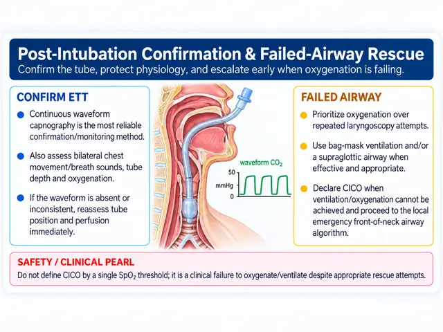
Rapid Sequence Intubation in the ICU — At-a-Glance Summary
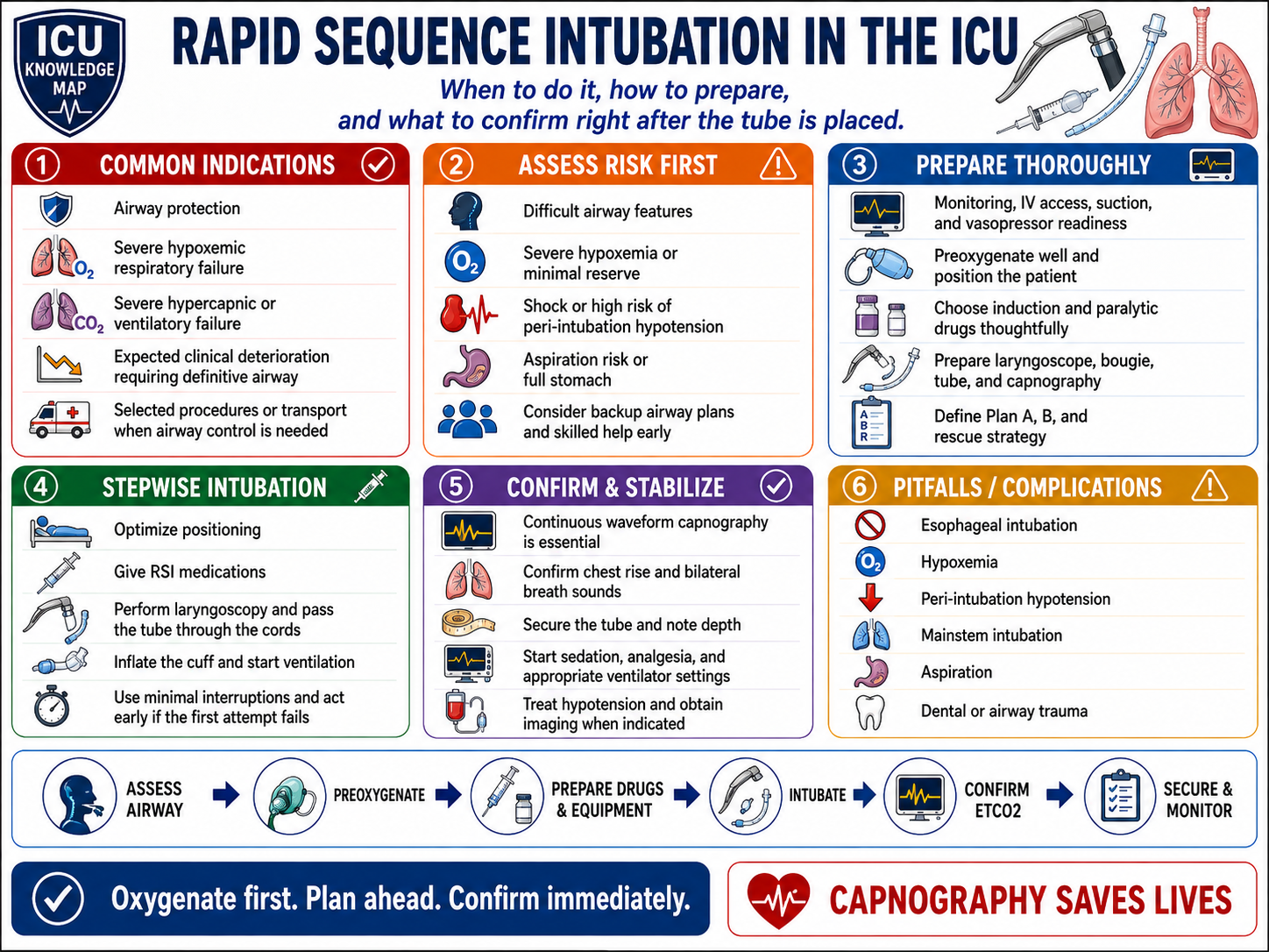
ICU Rapid Sequence Intubation — Safe Sequence
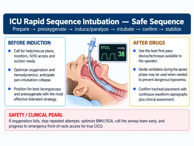
Post-Intubation Confirmation & Failed-Airway Rescue
Verify it on ICU Imaging
Practice recognizing correct device position, malposition and imaging complications after this procedure.
Indications & contraindications
Indications
- Failure to oxygenate or ventilate
- Loss or threatened loss of airway protection
- Expected clinical deterioration requiring controlled airway management
- Procedural or transport requirements when benefits outweigh risk
Contraindications / reasons to change strategy
- No absolute contraindication when an airway is immediately required
- Delay RSI when a safer awake or alternative airway strategy is indicated by predicted anatomic difficulty
- Severe physiologic instability is a reason to optimize first when time permits, not a reason to ignore the airway
Equipment checklist
Airway view & landmarks
Landmarks
- Epiglottis
- Arytenoids/interarytenoid notch
- True vocal cords
- Posterior glottic opening
- Tube passing through the cords
Visual target
Identify every listed landmark before advancing to the next irreversible step.
Danger zones
- Esophageal inlet posterior to larynx
- Trauma from repeated forceful attempts
- Loss of oxygen reserve during prolonged laryngoscopy
- Hemodynamic collapse after induction/positive pressure
Procedure video
Source-hosted videoEndotracheal Intubation via Video Laryngoscopy
This source uses its own hosted player. Open the original unchanged video at the source below.
Open procedure video ↗Preparation
- Assign roles and verbalize Plan A/B/C/D
- Optimize position; ramp obese patients and align external auditory meatus with sternal notch when appropriate
- Preoxygenate aggressively; consider HFNO/NIV/PEEP strategy according to physiology
- Treat hypotension before induction when possible and have vasopressor support running/ready
- Choose induction/paralytic doses based on shock, RV failure, asthma, intracranial pathology and other physiology
- Set the ventilator before the tube is placed
Step-by-step technique
Perform final team time-out: indication, predicted difficulty, drugs, rescue plan and hemodynamic plan
Preoxygenate to maximize safe apnea time; use apneic oxygenation when appropriate
Administer induction agent then rapid-onset neuromuscular blocker
Perform laryngoscopy and identify the glottis; optimize with positioning/external laryngeal manipulation or adjuncts rather than repeated identical attempts
Pass the tube through the cords to an appropriate depth; inflate cuff
Immediately attach continuous waveform capnography and ventilate
Secure the tube and reassess depth, bilateral ventilation and hemodynamics
Start analgesia/sedation promptly and transition to the pre-planned ventilator strategy
Hard stop points
- Stop and reoxygenate when saturation is falling or attempts are becoming prolonged
- Escalate immediately if ventilation/oxygenation cannot be restored
- Do not continue serial laryngoscopy attempts without changing the strategy
How to confirm success
- Sustained continuous waveform end-tidal CO2 is the primary confirmation method
- Bilateral chest rise and breath sounds support but do not replace capnography
- Check tube depth at teeth/lips and confirm position with imaging when clinically indicated
- Reconfirm after transfer, proning, major position change or unexplained deterioration
Complications & troubleshooting
Complications to watch for
Aftercare & documentation
- Document tube size/depth, device/grade/view, number of attempts and complications
- Order/perform appropriate post-intubation imaging
- Reassess sedation, analgesia, ventilator targets and VAP-prevention bundle
Test yourself before the bedside
Sequence recognition, hard-stop recall and complication response for RSI / Intubation. This is an educational self-check, not credentialing.
1. Sequence challenge
Score 0/5Perform final team time-out: indication, predicted difficulty, drugs, rescue plan and hemodynamic plan
According to this guide, what comes next?
2. Hard-stop recall
Before revealing them, name the situations in which you should stop, change strategy or escalate.
3. Complication response
Say your response first, then reveal the audited guide action.
Before / during / after
Interactive learning checklist for RSI / Intubation. Local policy, credentialing and patient-specific decisions take precedence.