Ultrasound-Guided Central Venous Catheter Insertion
Central venous access using real-time ultrasound, maximal sterile barrier precautions, wire safety and reliable confirmation before high-risk use.
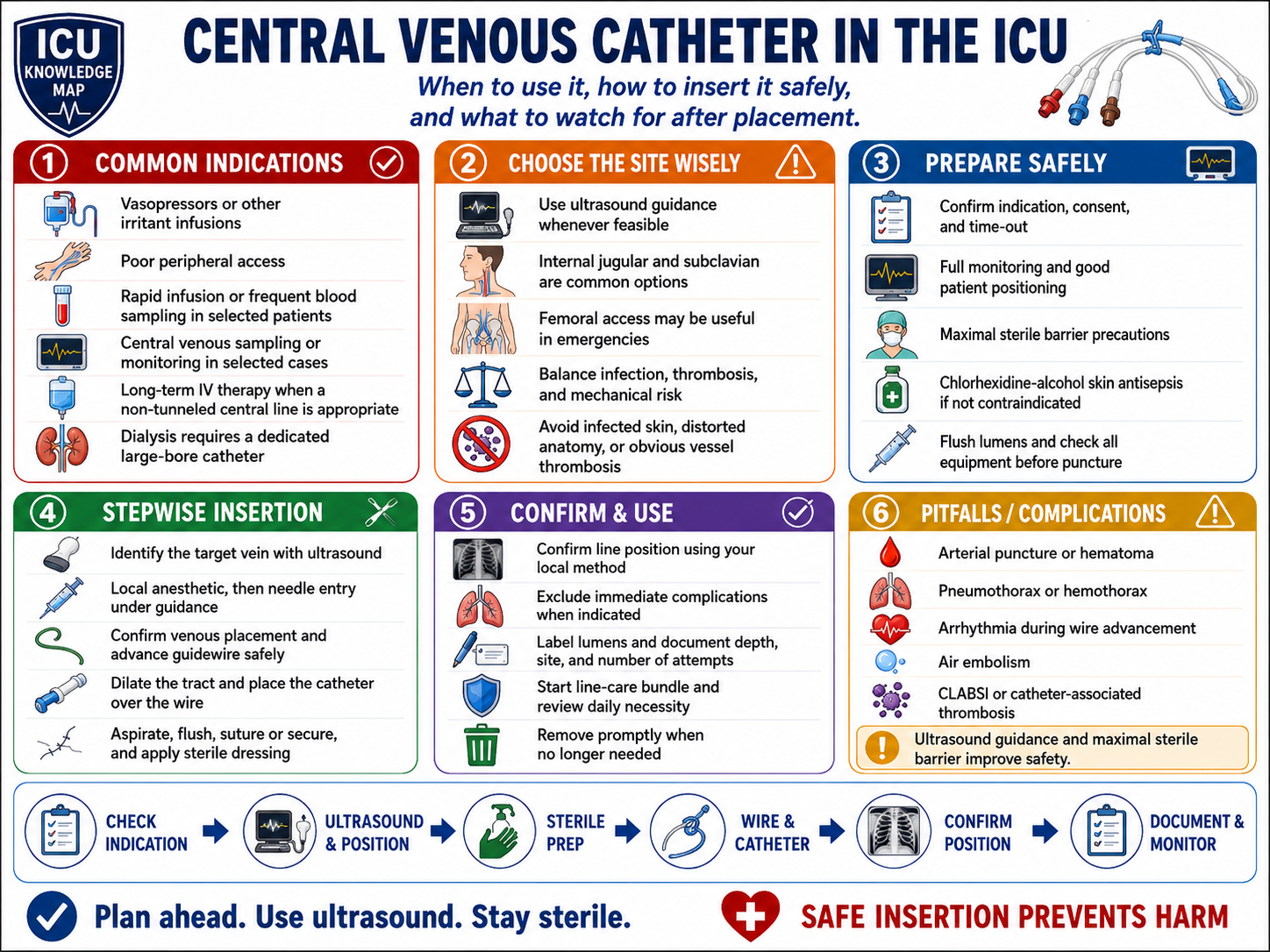
Central Venous Catheter in the ICU — At-a-Glance Summary
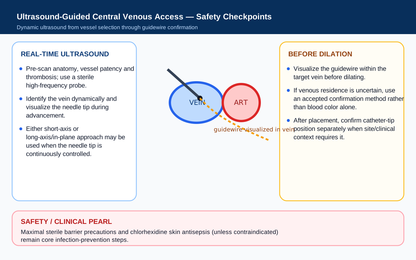
Ultrasound-Guided Central Venous Access — Safety Checkpoints
Verify it on ICU Imaging
Practice recognizing correct device position, malposition and imaging complications after this procedure.
Indications & contraindications
Indications
- Vasoactive infusions
- Reliable central access for selected irritant/hyperosmolar therapies
- Difficult peripheral access
- Hemodynamic monitoring when clinically useful
- Large-bore access or dialysis when an appropriate catheter is selected
Contraindications / reasons to change strategy
- Infection or burn at the insertion site
- Known thrombosis/occlusion of target vessel
- Uncorrected severe coagulopathy when a safer site/strategy exists
- Anatomy that cannot be safely visualized
Equipment checklist
Ultrasound vessel view
Landmarks
- Target vein and adjacent artery
- Compressibility and respiratory variation
- Needle tip entering the lumen
- Guidewire visualized within the vein before dilation
Ultrasound / visual target
- Use real-time needle-tip visualization
- Scan for thrombosis before puncture
- Confirm the wire remains intravascular before dilating
Danger zones
- Carotid/subclavian/femoral artery
- Pleura/lung apex for thoracic approaches
- Excessive wire insertion causing arrhythmia
- Dilating an unconfirmed vessel
Procedure video
External educational videoCentral Venous Catheter Insertion
Pre-insertion
Preparation, positioning, ultrasound assessment, sterile setup and equipment review (0:00–6:33).
Insertion
Needle/wire/catheter insertion and completion from 6:33 to the end.
Preparation
- Choose the site by balancing infection, mechanical-complication, thrombosis, bleeding and future dialysis-access risks. In adults, CDC guidance advises avoiding femoral CVC access when suitable alternatives exist; for a nontunneled CVC, subclavian access has lower infection risk than jugular/femoral access but must be balanced against pneumothorax/bleeding risk.
- Avoid subclavian access in hemodialysis patients and patients with advanced kidney disease when possible because central venous stenosis can compromise future permanent dialysis access.
- Use the minimum number of catheter lumens required for the clinical indication.
- Perform hand hygiene and maximal sterile barrier precautions: cap, mask, sterile gown, sterile gloves and a full-body sterile drape.
- Prepare clean skin with >0.5% chlorhexidine in alcohol unless contraindicated, and allow the antiseptic to dry according to the product instructions before puncture.
- Flush and prepare all lumens, caps and clamps before insertion.
- For adults undergoing CVC placement at a compressible site, the 2025 AABB/ICTMG platelet guideline recommends prophylactic platelet transfusion when the platelet count is <10 × 10^3/µL (conditional recommendation). Do not extrapolate this single threshold to noncompressible, large-bore or otherwise higher-risk access; individualize with the procedural context and local policy.
Step-by-step technique
Identify vein and artery with ultrasound and confirm patency
Infiltrate local anesthetic
Advance needle under real-time ultrasound until tip is clearly in the vein
Confirm venous blood/pressure when uncertainty exists
Advance guidewire without force and visualize it in the vessel
Make a small skin nick, then dilate only after wire position is certain
Advance catheter over the wire while maintaining control of the wire at all times
Remove wire, aspirate and flush every lumen
Secure catheter, apply sterile dressing and document depth
Hard stop points
- Never dilate without confident venous wire position
- Stop if resistance is encountered
- Treat sudden hypoxemia/hypotension after thoracic insertion as a possible procedural complication until excluded
How to confirm success
- Confirm venous placement before dilation when doubt exists
- Confirm final catheter tip/absence of pneumothorax using the locally accepted method: ultrasound, intracavitary ECG and/or chest radiography depending on site and circumstances
- Do not use for high-risk infusion if location remains uncertain
Complications & troubleshooting
Complications to watch for
Aftercare & documentation
- Daily line-necessity review
- Inspect site and dressing
- Use meticulous hub disinfection
- Remove promptly when no longer required
Test yourself before the bedside
Sequence recognition, hard-stop recall and complication response for Central Line. This is an educational self-check, not credentialing.
1. Sequence challenge
Score 0/5Identify vein and artery with ultrasound and confirm patency
According to this guide, what comes next?
2. Hard-stop recall
Before revealing them, name the situations in which you should stop, change strategy or escalate.
3. Complication response
Say your response first, then reveal the audited guide action.
Before / during / after
Interactive learning checklist for Central Line. Local policy, credentialing and patient-specific decisions take precedence.